Why do Dentists take x-rays

Why do dentists take x rays...
We will discuss the different types of x rays , the reasons why we use them them and why they are an essential tool in dentistry. When a dentist is looking at your teeth - he can only see the outer surfaces of your teeth and jaws.
X-rays allow your dentist to see what is happening INSIDE your teeth and gums. Dentist use these to evaluate your oral health and determine if there are any issues that require treatment. They are an electromagnetic wave of energy which can permeate materials that you cannot see through with the naked eye.
X-rays can be used to:
• Monitor growth and position of teeth in the child and adult
• Show decay that is not directly visible in the mouth and will also show the size and depth of a cavity. This will assist the dentist in deciding what type of filling will need to be used for that particular tooth.
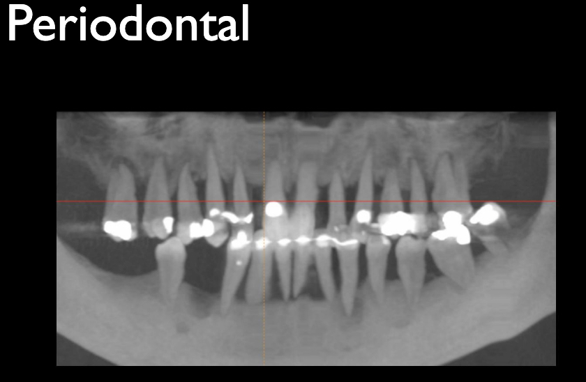
• Reveal the extent of bone loss in patients who are suffering from gum disease.
• Reveal an infection of the teeth, the gums and the bone.
• Reveal abnormal growths, such as a tumour.
• Determine the length of roots when carrying out a root canal treatment or extraction.
• Determine whether there is enough space in the mouth for new incoming teeth.
• Monitor the development of wisdom teeth.
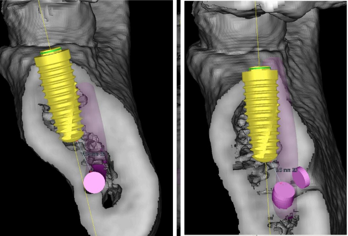
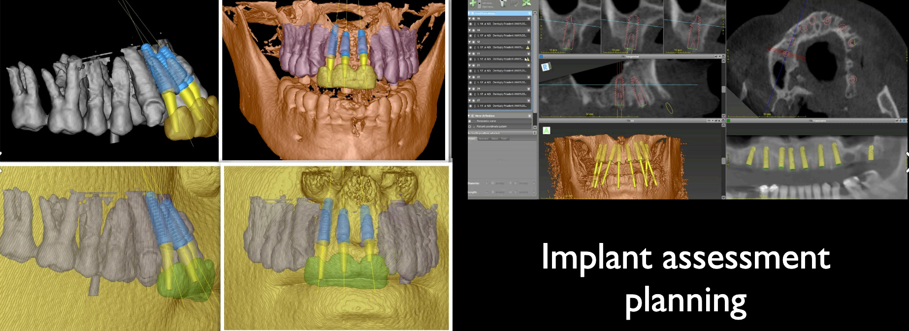
Implant assessment and sizing the correct implant within the available bone
.
If you are a new patient, the dentist will normally take x-rays at the initial visit to be able to assess your mouth and determine whether any treatment is necessary. After this, it is considered good practice to have x-rays taken probably every 1-2 years. However, this may vary depending on the dental history of a patient, such as whether they have poor oral hygiene and whether they continue to get new cavities. It will also depend on the age and current condition of the patient’s mouth. It may therefore be the case that x-rays are required more frequently and your dentist should assess this based on individual circumstances.
Evolution of X-rays
Despite any reservations, real or perceived, x-ray imaging was one of the most important developments in the history of dentistry. Dentists often need to treats issues below the gumline. Without the aid of x-rays, they would be forced to make an incision and turn something simple into an invasive procedure.
The x-ray was first discovered in 1895 by Wilhelm Röntgen and in 1896 the first dental x-ray was taken by C. Edmund Kells in New Orleans. Over the decades, the technology advanced to feature faster film speed, better quality images and increased comfort for patients. Eventually, x-rays were established as an acceptable and necessary tool for dentists. By the 1950s, they were in regular use and today are present in virtually every dental clinic
Dentists have several options with regards to the type of dental x-rays they can perform. Some of the more common are:
A Bitewing, a technique where the patient bites a specific type of paper as the x-rays are taken. This allows the dentist to see how the crowns of the teeth match up and to diagnose gum disease and cavities. Its a bit like a "Portrait" of the teeth
A Panoramic is a x-ray that rotates around the face. This helps the dentist examine wisdom teeth and jaw abnormalities. This is the "group" photo at the wedding that lets you see who is threr, who is not, and who is related to who and looking well!
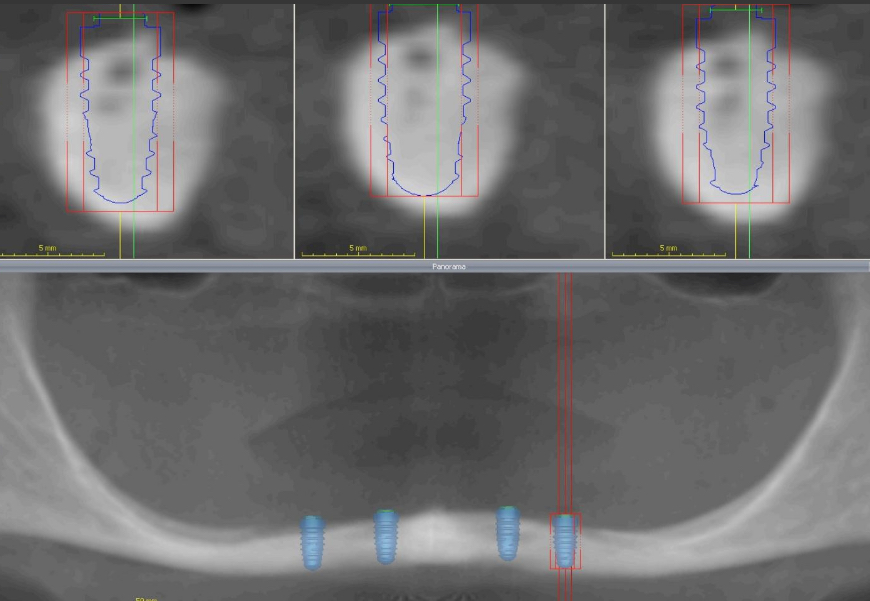
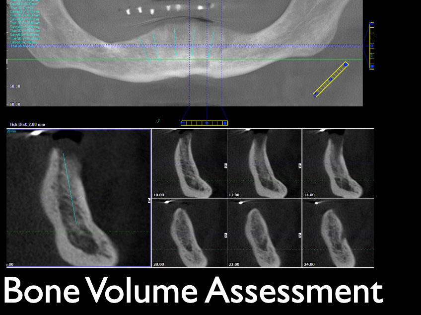
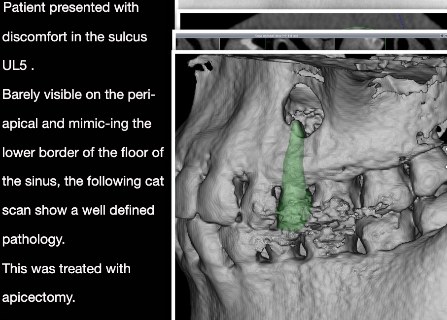
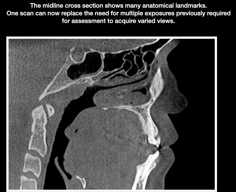
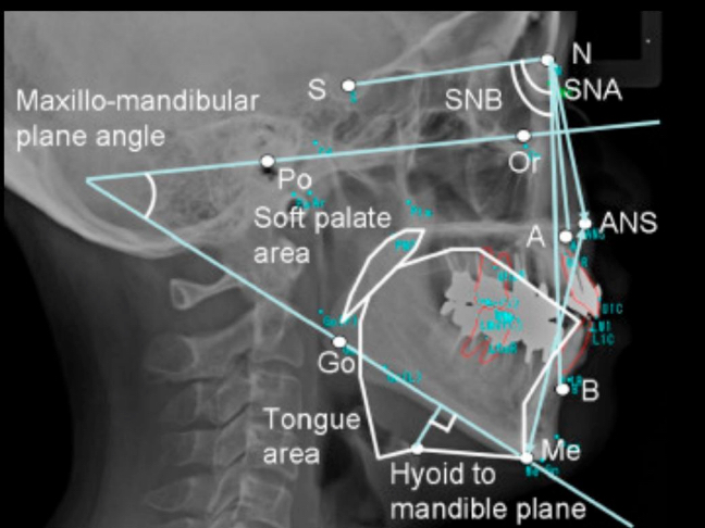
A Cat scan or CBCT ( Cone Beam Computerised Tomography ) or 3 dimensional cat scan allows the dentist to see a three dimensional view of your teeth and jaws. They can look through cross sections and measure distances with unprecedented accuracies to assess bone levels for implant placement, the degree and extent of infections and pathologies, - the presence of nerves and proximity to wisdom teeth, the position of teeth and roots for orthodontic assessment......
We always recommend speaking with your dentist or dental hygienist about your x-rays. It’s never a bad idea to ask what x-rays are being taken and for what reasons, especially if it will put you at ease.
Your dentist will always minimise the x rays to those that will give suitable clinical information.
Digital X-rays
Digital x-rays were an enormous innovation in the field of dentistry that reduced the dosage and improved the quality and ease of imaging, and not just because it’s a safer method. It helps dentists improve their diagnosis and start treatment quicker because they can clearly and quickly identify issues.
The more information we have about a patient’s teeth and gums, the more likely we will be able to pinpoint any problems and provide the highest standard of care.
In addition to providing quicker insights into a patient’s case while exposing them to minimal radiation, digital x-rays allow the dentist to control the contrast, colour and brightness. This control is essential to providing our patients with the best oral care.
Below are a collection of x ray images we might see in a dental practice.
Leave a Comment